Methods paper accepted in IUCr
The preprint of our new methods paper is online
We are pleased to announce our first method paper actually published on BioRxiv, entitled "Revisiting Sodium Phosphotungstate and Ammonium Molybdate as Non-Radioactive Negative Staining Agents for Single Particle Analysis."
In this study, we have successfully replaced uranyl-based stains with sodium phosphotungstate or ammonium molybdate for negative staining electron microscopy. Our tests with apoferritin demonstrated that these non-radioactive stains, when combined with a straightforward on-grid fixation step, produce images comparable to those obtained with uranyl formate.
Further experiments with β-galactosidase confirmed that these new stains are also effective for single particle analysis, yielding results virtually indistinguishable from those with uranyl formate. Since sodium phosphotungstate and ammonium molybdate are non-radioactive, they avoid the stringent handling restrictions associated with uranyl-based stains.This not only reduces costs but also broadens access to decentralized sample grid preparation immediately after purification, benefiting a wider range of scientists in the cryo-EM community.
Read the full article on BioRxiv for a detailed account of our findings and a step-by-step description of negative staining with phosphotungstate or ammonium molybdate.

Direct Electron Detectors: an interplay between the two available modes

An article written by our lab student Malika Askarova about the results of her lab module
With the development of direct electron detectors (DED), electron microscopy has seen a tremendous leap in the resolution achievable. Unlike their conventional counterpart – the charge coupled devices (CCD) that converts electrons to photons and thus produce an image – DEDs operate by directly receiving incident electrons, which eliminates parts of the noise and reduces loss of signal due to electron-to-photon conversion.
At the StruBiTEM facility, we employ a Falcon III direct electron detection camera (Thermo Fisher Scientific, MA, USA) that allows two modes of data acquisition: integrating and counting. In integrating mode, the signal arising from incident electrons gets integrated, and the signal contains contributions from a large number of incident electrons. Due to Landau distribution, each electron depositing different amounts of energy inevitably causes Landau noise, leading to a lower signal-to-noise ratio (SNR) in the image. In electron counting mode, Landau noise is fully eliminated, since individual electrons hitting the detector are counted as distinct incidents (1 or 0), therefore there is no energy variability.
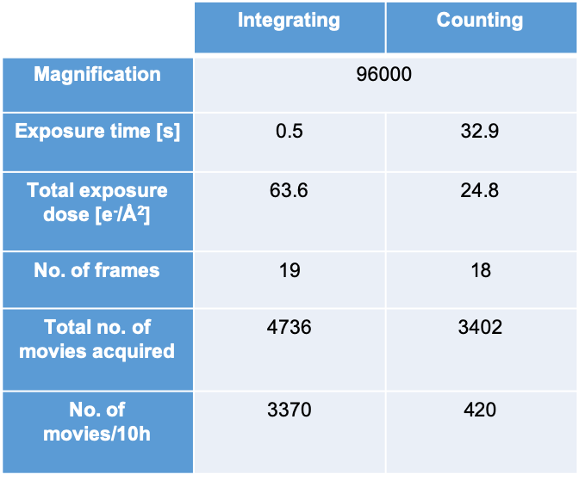
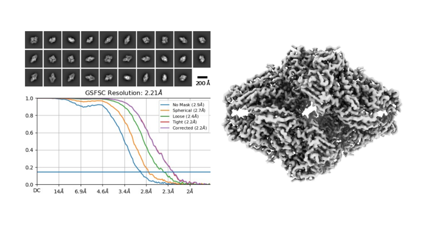
When acquiring 1000 micrographs in each of the modes, we observe a slight advantage of the counting mode over the integrating mode in resolution, with 2.17 Å resolution in counting mode vs. 2.31 Å resolution in integration mode (Figure 1). When comparing the 2D classes generated in both modes, those of electron counting mode subjectively look better, sharper and with less background noise (Figure 1a and 1b).
Concerning the throughput, integrating mode produces more images than counting mode. For the total exposure dose of 63.6 e-/Å2 in integration mode, the exposure time was only 0.5 s. The total dose in counting mode was 24.8 e-/Å2 and therefore the acquisition time is 32.9 s. Table 1 summarizes the data for both of the modes.
Taking the different exposure times in account and keeping in mind that the most limiting factor at the Krios is acquisition time, a fair comparison would be to acquire data for 10 h in each mode and compare the resulting resolution. Throughout an exposure of 10 hours, 3370 movies were acquired in integrating mode, whereas 420 were taken in counting mode. Here, the resolutions achieved were 2.48 Å for integrating mode and 2.21 Å for counting mode.
The question arising is whether to use integrating mode for faster acquisition or counting mode for acquiring data of higher quality. Even when time is the limiting factor, the counting mode was superior. The data acquired in 10 h resulted in higher resolution in counting mode, even though 8 times less data were produced. For small proteins, the higher resolution and the better SNR in counting mode will be helpful, especially for the particle alignment, while for larger particles with high symmetry that are easy to align, integrating mode might be the choice, since it allows fast acquisition of large numbers of particles.
Ensuring constant quality
February 9th 2022
Also our second test sample, the tetrameric beta-galactosidase with a total molecular weight of 465 kDa, easily reached near-atomic resolution. Indeed, with a resolution of 2.2 Å we are close to the current record for this protein which is at 1.8 Å. For our dataset, the whole process of plunge freezing, grid screening and data acquisition of the 5000 frame movies used was completed within a single day.
We will continue using test specimens, such as beta-galactosidase or apoferritin, to ensure that our microscopes are always working at peak condition in order to guarantee optimal measurements for our users.
Connection established
February 8th 2022
One aim of our facility is to make use of cryo-EM as a scientific tool as easy as possible for our users. One aspect in this regard is that we we strive to centralise computation to prevent that each and every workgroup has to buy, install and maintain their individual workstation. With the RRZK we have the ideal partner for this - however we still need to get the data from our microscopes into the compute cluster.
As of today we have established a direct glasfiber connection to CHEOPS that will allow our users to transfer their data directly to the compute cluster and use the central hardware and software resources to drive their science projects. First tests have shown that we can sustain transfer rates of 75 MB/sec, allowing us to transfer full-size frame movies in less than 10 seconds. This high transfer rate will enable us to use the computational resources available at CHEOPS to establish a pipeline allowing us to reconstruct 3D structure in real-time while data acquisition is still running. Currently, this is still in the development stage, but we will hopefully soon be able to post further news on this.
Krios passed its trial by fire
October 15th 2021
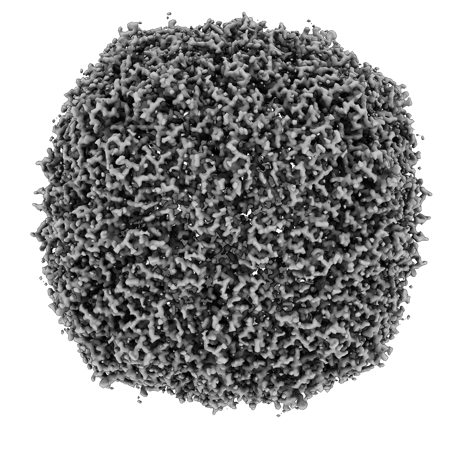
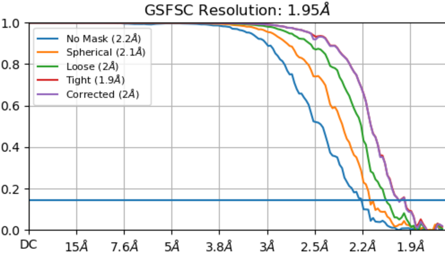
We can now confirm that the Titan Krios is performing exceptionally well and has generated its first near-atomic resolution structure. Using apoferritin as a test specimen we could reach a resolution of 1.95 Å using very basic experimental conditions: standard copper grids and integration instead of counting images. In total we only included 2700 frame movies into this dataset, allowing us to finish the processing within two days after completing the dataset acquisition.